What is AVM
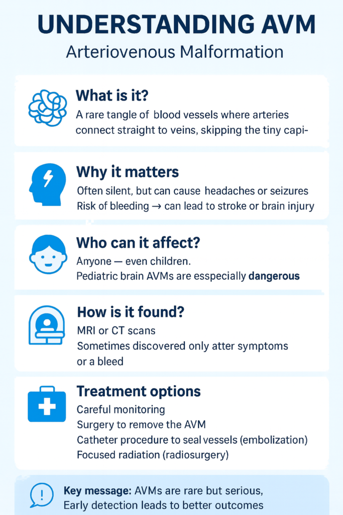
A pediatric arteriovenous malformation (AVM) is a rare condition where a cluster of arteries connects directly to veins without the normal tiny vessels, called capillaries, in between. This abnormal shortcut creates fragile, high-pressure blood flow that puts children at greater risk of brain hemorrhage or stroke. Unlike adults, children often present with larger and more complex AVMs, which tend to behave more aggressively and carry a higher lifetime risk of rupture simply because of the many years ahead of them.
Most children with AVMs come to medical attention after a sudden event. In fact, the most common first sign in pediatric patients is a bleed (hemorrhage), which can cause severe headaches, vomiting, loss of consciousness, or even life-threatening stroke. Other children may experience seizures, persistent headaches, or subtle neurological symptoms like weakness, vision problems, or learning difficulties. Some AVMs remain silent until a rupture occurs, making early detection especially challenging.
Diagnosis and treatment
Doctors use imaging like MRI and MRA to locate AVMs, while CT is reserved mainly for emergencies involving bleeding. For the most precise detail before treatment, cerebral angiography (DSA) reveals the exact vessel anatomy.
Because children with AVMs face a lifetime risk of rupture, treatment—rather than mere observation—is usually recommended. Microsurgical resection is preferred when the AVM is accessible. Endovascular embolization blocks abnormal vessels via a catheter. Radiosurgery uses focused beams of radiation to shrink AVMs over 2–3 years, though it’s used cautiously in very young children.
Often, a combination of treatments is used. Outcomes have improved with modern techniques, but long-term follow-up is essential: recurrence or new vessel growth is possible even after apparent obliteration. With early diagnosis, careful treatment, and ongoing monitoring, many children can lead healthy lives.
Our Focus
Research
Advancements in early detection of pediatric arteriovenous malformations (AVMs) are crucial for improving patient outcomes. A recent study by UCSF neurosurgery resident Dr. Alex Lu and Chief of Pediatric Neurosurgery Dr. Nalin Gupta analyzed a 24-year database of pediatric AVM cases. Their research, published in the Journal of Neurosurgery: Pediatrics, compared the characteristics and management of incidental versus symptomatic unruptured AVMs in children. UCSF Neurosurgery
The study revealed that incidental AVMs were generally smaller and more accessible surgically than symptomatic ones. Incidental AVMs smaller than 3cm were successfully treated with surgery. The research also suggested that pediatric AVMs may grow over time without treatment. These findings underscore the importance of early identification and intervention.
Early screening
A simple brain scan such as an MRI can reveal these dangerous tangles of blood vessels before they cause irreversible harm. Making MRI or another safe form of neuroimaging mandatory for children, at least once during early development, would allow doctors to identify hidden AVMs, guide timely treatment, and prevent strokes or life-threatening hemorrhages. Just as we screen children for hearing or vision problems, routine scanning for AVMs could save lives, reduce long-term disability, and offer families peace of mind.
With your generous support, we aim to fund research initiatives like this to develop strategies for detecting and treating AVMs during a child’s development, potentially preventing future complications.